
Document Type : Research Paper
INTRODUCTION
Metal-oxide nanostructures continue to attract sustained interest in catalytic, optical and biological behaviors because of their high surface-to-volume ratio, tunable defect chemistry and rich surface acidity. Among them, zirconium dioxide (ZrO2), sometimes simply called zirconia, occupies a particular place. The material is hard, chemically inert, thermally robust (melting point ≈ 2710 °C) and biocompatible, which is why it has long been used in dental restorations, thermal-barrier coatings, oxygen sensors and solid-oxide fuel cells [1-3]. At the nanoscale, however, the chemistry shifts: surface oxygen vacancies become more numerous, the band-gap edges sharpen, and the material develops antibacterial activity [4, 5]. Several physical and chemical routes have been used to obtain ZrO2 nanoparticles, including sol–gel processing, hydrothermal treatment, co-precipitation, microwave-assisted synthesis and laser ablation [6-8]. While each technique has its advantages, the disadvantages are similar: high temperature, high cost of surfactant, hazardous reducing agents (such as hydrazine and NaBH4), and/or organic solvents that are difficult to dispose of. However, concerns have brought the field to the “green” synthesis where the use of the conventional reagents is replaced by plant extracts. As a whole, phytochemicals like flavonoids, tannins, terpenoids and phenolic acids are not only reducing agents but also natural capping/stabilizing molecules, in which the conversion of metal to oxides takes place in water, under moderate temperature and by-products are biodegradable [9, 10].
Several plants were used for bio-mediated synthesis of ZrO2. All of these can be given as examples: Aloe vera gel [4, 11], extract of the leaves of Eucalyptus globulus [12] and of Wrightia tinctoria [13] and Lagerstroemia speciosa [14]. Among less explored candidates is mint (Mentha spicata L.), herb whose leaves are particularly rich in rosmarinic acid, menthol, carvone and methylated flavonoids. Its phytochemical profile makes mint attractive as a multi-functional bio-template: the polyphenols reduce Zr4+, the carbonyl groups bind to nascent particle surfaces, and the menthol fraction may slow grain growth during calcination. To the best of our knowledge, comparatively few systematic studies have correlated the structural, morphological and antibacterial behaviour of mint-derived ZrO2. The present work was undertaken with three objectives. First, to demonstrate a simple aqueous green-synthesis protocol for ZrO2 using mint-leaf extract and Zr(NO3)4 as precursor. Second, to characterize the calcined powder by X-ray diffraction (XRD), field-emission scanning electron microscopy (FE-SEM) and energy-dispersive X-ray spectroscopy (EDX) in order to verify phase, morphology and chemical purity. Third, to evaluate the antibacterial response of the prepared nanopowder against one Gram-positive (S. aureus) and one Gram-negative (E. coli) bacterium, while comparing the influence of two precursor molarities (0.5 M and 1 M) on the activity.
MATERIALS AND METHODS
Chemicals and biological materials
Zirconium nitrate pentahydrate, Zr(NO3)4.5H2O (analytical grade, ≥ 99 %), and sodium hydroxide pellets (NaOH, ≥ 98 %) were used as received without further purification. Distilled water was used throughout experiments. Fresh mint (Mentha spicata L.) leaves were obtained from a local market, and the bacterial strains S. aureus (ATCC 25923) and E. coli (ATCC 25922) were supplied by the microbiology unit of the College of Science, University of Wasit.
Preparation of the mint-leaf extract
Approximately 25.0 g of fresh, undamaged mint leaves were rinsed several times with tap water and then with distilled water to remove dust and surface contaminants. The leaves were chopped into small pieces and immersed in 250 mL of distilled water held at 60 °C for 30 min under gentle magnetic stirring. The greenish extract was allowed to cool to room temperature and was filtered through Whatman No. 1 paper. The clear filtrate was kept refrigerated at 4 °C and used within 48 h.
Synthesis of ZrO₂ nanoparticles
A solution of Zr(NO3)4.5H2O with a concentration of 1.0 M in distilled water was prepared by dissolving the corresponding mass of the salt in 50 mL of distilled water, while a second solution of the same salt was prepared with a concentration of 0.5 M in the same way, for the study of the dependence of molarity. The precursor solution was then drop-wise added to the mint extract at 60 °C with continuous magnetic stirring, such that the ratio of extract to precursor solution was maintained at about 1:1. NaOH solution was added drop-wise after 30 min of mixing until the pH value reached 9 ± 0.2, with almost immediate appearance of a milky-white precipitate. The stirring was continued for another hour to insure complete precipitation. The slurry was placed in centrifuge tubes (4000 rpm, 10 min) and the supernatant was removed. The solid collected was washed with distilled water/ethanol and dried overnight in a hot-air oven at 80 °C. Finally, the dried powder was converted into a fine white powder of ZrO2 nanoparticles by calcining it in a muffle furnace at 600 °C, for 2 h with a heating rate of about 5 °C min–1. A flow chart of the overall process is given in Fig. 1.
Antibacterial assay
The biocidal activity of the calcined ZrO2 nanopowders (0.5 M and 1.0 M batches, both pH 9, calcined at 600 °C) was assessed using the agar-well diffusion technique. Mueller–Hinton agar plates were inoculated with 0.1 mL of fresh bacterial suspension (≈ 106 CFU mL–1) and spread uniformly with a sterile glass rod. Wells of 6 mm diameter were punched aseptically and each was filled with 50 µL of an aqueous nanoparticle suspension (50 µg mL–1). The plates were incubated at 37 °C for 24 h, and the diameter of the clear zone surrounding each well was measured using a digital vernier caliper. Each test was performed in triplicate, and the mean value was reported.
Characterization techniques
The crystalline phase and crystallite size of the calcined powder were investigated using a powder X-ray diffractometer with Cu-Kα radiation (λ = 1.5406 Å) over the 2θ range 10°–80°. Surface morphology was studied with a TESCAN MIRA3 field-emission scanning electron microscope operating at 20 kV, using the in-beam secondary-electron detector. Elemental composition and elemental mapping were obtained from the EDX detector coupled to the same instrument. The antibacterial behavior was tested by the agar-well diffusion method.
RESULTS AND DISCUSSION
XRD analysis
The XRD pattern of the ZrO2 powder calcined at 600 °C is shown in Fig. 2. The diffractogram displays a set of sharp, well-defined Bragg peaks superimposed on a low and almost flat background, which indicates that the prepared powder is reasonably crystalline despite the moderate calcination temperature. All the reflections could be indexed to the cubic phase of ZrO2, in good agreement with JCPDS Card No. 96-152-1754. No additional peaks belonging to monoclinic or tetragonal ZrO2, to ZrO2·xH2O or to carbonaceous residues from the mint extract were observed, suggesting that calcination at 600 °C was sufficient to drive off the organic capping layer while stabilizing the cubic polymorph. Although the cubic phase of zirconia is normally unstable below ~2370 °C, several authors have shown that nanocrystalline c-ZrO2 can persist at room temperature when the crystallite size is below a critical limit (≈30 nm) [15, 16]. The present results fall comfortably within this size window. The three strongest reflections were located at 2θ = 30.290°, 50.511° and 60.044°, corresponding to the (111), (202) and (311) planes, respectively. The experimentally measured d-spacings (2.948, 1.805 and 1.539 Å) match the standard values (2.938, 1.799 and 1.534 Å) within ≤ 0.4 %, which lies within the resolution limit of the diffractometer and confirms the structural assignment.
The average crystallite size, D, was estimated from the Scherrer equation:
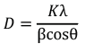
where K (≈0.9) is the shape factor, λ is the X-ray wavelength, β is the full-width-at-half-maximum of the peak (in radians) and θ is the Bragg angle. The values obtained for the three planes were 16.53, 16.6 and 14.6 nm, giving an average crystallite size of 15.91 nm (Table 1). The slight reduction in size at higher 2θ (the (311) plane) is consistent with anisotropic growth and with the typical broadening observed for high-index reflections in nanocrystalline oxides.
FE-SEM morphology
Representative FE-SEM images recorded at different magnifications (Fig. 3) reveal that the calcined ZrO2 powder consists of nearly spherical to quasi-spherical primary particles. Some clustering can be seen, particularly at lower magnification, where the particles tend to form loose aggregates of a few hundred nanometers. This agglomeration is not unusual for oxide nanopowders calcined at 600 °C and is generally attributed to the high surface energy of the nanocrystallites combined with the partial sintering of neck regions during heat treatment [17]. At scale of 200 nm, the individual grains become visible, and most of them lie in the 15–30 nm range, in good qualitative agreement with the XRD-derived crystallite size of ≈15.9 nm. The somewhat larger apparent grain size in SEM relative to XRD is consistent with the well-known fact that XRD reports the size of coherently diffracting domains, while SEM detects the whole particle, which may contain more than one such domain.
EDX and elemental mapping
The EDX spectrum (Fig. 4) shows two strong peaks corresponding to Zr (Kα ≈ 2.04 keV, with weaker L lines below 2 keV) and O (Kα ≈ 0.52 keV). No peaks associated with carbon, nitrogen or sodium were detected, demonstrating that the washing protocol effectively removed nitrate and sodium residues, and that calcination eliminated the organic capping layer. The quantitative output gave 65.4 wt % Zr and 34.6 wt % O (corresponding to 56.5 at % Zr and 43.5 at % O). The Zr:O atomic ratio of about 1:0.77 is below the stoichiometric value expected for ZrO2 (1:2); this slight oxygen deficiency is characteristic of nanocrystalline ZrO2 and is usually associated with surface oxygen vacancies, which are also responsible for the cubic-phase stabilization observed by XRD [18]. The EDX elemental maps confirmed a uniform spatial distribution of Zr and O over the sample area, with no segregation or localized clustering.
Antibacterial activity
The anti-bacterial effect of the green synthesized ZrO2 nanoparticles is summarized in Table 2 and illustrated in Fig. 5. The clear, well-defined inhibition zones around the wells that had the nano powder showed both bacterial strains inhibited growth. In the case of the control wells without ZrO2, the inhibition zone was 0 mm. These data show that the effect is concentration-dependent: that is, when the initial molarity of the precursor (the more densely packed the nanoparticles in the well) is higher, the zone of inhibition is larger, and this happens more strongly in the case of E. coli (Δ = 4 mm) than S. aureus (Δ = 1 mm). Second, S. aureus (Gram-positive) was systematically more sensitive than E. coli (Gram-negative). This pattern is consistent with several reports on metal-oxide nanoparticles [19, 20] and is normally attributed to differences in the cell-wall architecture: the thick but relatively permeable peptidoglycan layer of Gram-positive bacteria allows easier ingress of nanoparticles and reactive oxygen species, while the outer lipopolysaccharide membrane of Gram-negative bacteria functions as an additional diffusion barrier.
The biocidal mechanism of ZrO2 nanoparticles is generally accepted to involve a combination of three pathways: (i) direct membrane disruption by sharp-edged nanoparticles and electrostatic interaction between the slightly positive nanoparticle surface and the negatively charged bacterial wall; (ii) generation of reactive oxygen species (•OH, O2•−, H2O2) at oxygen-vacancy sites of the cubic lattice, which oxidize lipids, proteins and DNA; and (iii) leaching of Zr4+ ions, and can interfere with intracellular enzymatic processes [21, 22]. The high oxygen-vacancy density inferred from the sub-stoichiometric EDX composition supports the second pathway as a dominant contribution in the present sample.
CONCLUSION
A simple, low-cost, and environmentally benign route for ZrO2 nanoparticles has been demonstrated using fresh mint (Mentha spicata) leaf extract as a multi-functional bio-template. Calcination at 600 °C produced a phase-pure cubic ZrO2 powder with an average crystallite size of 15.91 nm, as confirmed by XRD. FE-SEM revealed quasi-spherical primary particles in the 15–30 nm range, while EDX confirmed elemental purity and a slight oxygen sub-stoichiometry consistent with the stabilization of the cubic polymorph by surface vacancies. The agar-well diffusion test showed that the nanoparticles are biologically active against both S. aureus and E. coli, with maximum inhibition zones of 27 mm and 25 mm, respectively, at 1.0 M precursor concentration. The activity is dose-dependent and stronger against the Gram-positive strain, in line with the cell-wall-permeability argument. Overall, the results indicate that mint-mediated green synthesis is a viable, scalable alternative to conventional chemical routes for the production of antibacterial ZrO2 nanoparticles, with potential applications in wound dressings, water disinfection, and antimicrobial coatings.
CONFLICT OF INTEREST
The authors declare that there is no conflict of interests regarding the publication of this manuscript.
