
Document Type : Research Paper
INTRODUCTION
Alzheimer’s disease (AD) is the most common form of dementia and is a leading cause of cognitive decline, disability and dependence in older people [1]. It is a progressive neurodegenerative condition that markedly affects the elderly demographic and is chiefly defined by cognitive decline, memory deficits, and diminished daily functioning [2]. The neuropathological features of AD are defined by two main hallmarks, namely the extracellular accumulation of Aβ peptides leads to the formation of diffuse and neurotic plaques, alongside the intracellular buildup of neurofibrillary tangles (NFTs) comprised of hyperphosphorylated tau proteins. [3][4]. These pathological hallmarks are distributed throughout the brain and cause neuronal atrophy and synaptic loss, leading to neurodegeneration [4,5].
Besides these pathological changes, glutamatergic dysfunction and glutamate-mediated excitotoxicity have also been involved in neuronal damage and cognitive impairment in AD [6]. Notwithstanding advancements in comprehending genetic risk determinants and clinical presentations, the primary cause of AD continues to be unidentified. This ambiguity underscores the necessity for therapeutic approaches that target both symptom alleviation and potential disease-modifying processes. Nonetheless, existing treatments primarily focus on alleviating symptoms rather than addressing the root cause. The U.S. Food and Drug Administration (FDA) has sanctioned four medications: donepezil, rivastigmine, galantamine, and memantine. Cholinesterase inhibitors (donepezil, rivastigmine, and galantamine) are thought to improve cholinergic function, while memantine, an NMDA receptor antagonist, regulates glutamatergic activity [7]. NMDA receptor blocker Memantine This is employed to mitigate glutamate-associated excitotoxicity in moderate to severe instances of Alzheimer’s disease [8]. Furthermore, N-methyl-D-aspartate receptor antagonists are exclusively taken orally; however, approximately 57–82% of the unaltered medication is excreted via urine [9]. Therefore, delivery of drugs across the blood brain barrier (BBB) is one of the major challenges in AD therapy [10]. To overcome these limitations, nanoparticle (NP)-based systems for drug delivery have been developed as a promising approach. Nanoparticle based systems for drug delivery are coming up as promising strategies for improving drug stability, brain-directed delivery and hopefully, transport across the BBB[11-13]. Thus, loading anti-Alzheimer’s drugs in polymeric nanoparticles is a viable strategy for improving therapeutic effectiveness. Most polymeric NPs are biocompatible but viability as drug delivery vehicles in the CNS depends on biodegradability, physicochemical properties, stability and ability to cross biological barriers [13].
With the swift progress in nanotechnology in recent years, integrating bioactive elements into polymer substances for The regulated therapeutic release has emerged as a crucial area of investigation. Enclosing bioactive compounds within pharmaceutical delivery mechanisms Drug delivery systems (DDSs) serve as an effective method to safeguard these components from deterioration in extreme conditions, mitigate adverse effects, and enhance. Solubility, attain regulated release, and enhance biocompatibility [14,15].
NP-based systems represent a flexible and encouraging strategy to address the challenges posed by the blood-brain barrier in Alzheimer’s disease therapy. Nanoparticles have the potential to enhance treatment results for individuals afflicted with Alzheimer’s disease [16]. by improving drug stability, targeting ability, and penetration into the brain. In this perspective, natural polymers have attracted much attention as drug carriers. Their inherent advantages such as biocompatibility, controlled enzymatic degradation, specific biomolecular interactions, and ease of chemical modification have been well documented in the literature [17]. In biomedical applications, such as pharmaceuticals and tissue regeneration [18]. natural polymers derived from microorganisms, algae, plants, or animals have been widely used. Natural polymers are preferred to synthetic polymers from petroleum because of their excellent biocompatibility, natural degradability and sustainable origin [19,20]. Chitosan is insoluble in numerous solvents; however, it dissolves in dilute organic acids such as acetic, succinic, formic, and lactic acids with a pH below 6.0, owing to the primary amino groups on its backbone that generate a potent base(12). The production of blended polymers has emerged as a practical technique for imparting specific characteristics to polymeric materials for real-world use [21,22]. Chitosan (CS) is a linear cationic polysaccharide obtained by partial deacetylation of chitin and composed of β-(1→4)-linked glucosamine and N-acetylglucosamine units. This positive charge enables chitosan to interact electrostatically with negatively charged biological membranes and biomolecules under acidic conditions (when the amino groups are protonated) [23,24]. Chitosan-derived materials are highly regarded in biomedical fields due to their beneficial physicochemical and biological characteristics. Moreover, the net positive ionic charges of chitosan facilitate interactions with negatively charged surfaces, thereby validating its application in the creation of nanostructured biomedical materials [21].CS-derived nanocarriers have been investigated as potential platforms for brain-targeted drug delivery due to their biocompatibility, biodegradability and potential for enhanced drug transport to the CNS [25].
Importantly, the protonated amino groups of CS can interact electrostatically with anionic species such as phosphate groups. This property was used to prepare CS-sodium tripolyphosphate (TPP) nano particles by ionic gelation. The procedure relies on the electrostatic attraction between the positively charged amino groups of chitosan and the negatively charged phosphate groups of TPP, resulting in the creation of nanoparticles in mild aqueous environments [26,27]. Polymeric nanoparticles can encapsulate or associate with therapeutic agents and offer the possibility of improved drug stability, loading and controlled or sustained release [28]. Boswellia species, also known as frankincense, are rich in bioactive compounds including boswellic acids, which have demonstrated anti-inflammatory, antioxidant and neuroprotective effects. Experimental and clinical studies have indicated possible benefits of Boswellia-derived preparations in cognitive impairment and Alzheimer’s disease, but the mechanisms and therapeutic efficacy need further study [29,30]. The rapid development of nanoparticle-based strategies for AD therapy is paralleled with many reported formulations aimed at delivering single therapeutic agents. So far, few studies have been reported about the combination of conventional anti-AD drugs and bioactive natural products in a single biodegradable nanocarrier. This approach could provide the opportunity to combine the complementary pharmacological activities together with the enhancement of the drug localization and release features. Therefore, the current study is aimed to prepare and characterize chitosan-based nanoparticles by ionic gelation for co-delivery of memantine and natural extract of boswellia. The developed nanoparticles were characterized for particle size, polydispersity index, zeta potential, morphology, encapsulation efficiency and in vitro drug release behavior. The objective of this work was to develop a natural based drug delivery system, based on a natural polymer and a natural bioactive compound, for possible application in Alzheimer’s disease therapy.
MATERIALS AND METHODS
Chitosan (nano grade, average particle size (APS)<80 nm, viscosity<100 cP, D.A.C.<90%) was purchased from Xi’an Rongsheng Biotechnology Co., Ltd., China and used as received without any further purification. The glacial acetic acid (extra pure grade; molecular weight 60.05 g / mol) was procured from Thomas Baker Chemicals, India. Memantine hydrochloride (≥98%, MW = 215.76 g mol−1, CAS No. 41100-52-1) was purchased from Macklin China and was used as received. Sodium tripolyphosphate (TPP; Na5P3O10; molecular weight 367.86 g/mol) was purchased from Tianjin Dingshengxin Chemical Industry Co. Ltd, China and used as ionic cross-linker for the preparation of the nanoparticles. The extraction solvent was 70% (v/v) Ethanol (99%, v/v) prepared by diluting Ethanol with distilled water. The resin of Boswellia sacra (Omani frankincense) was obtained from a commercial source in the Sultanate of Oman. The experiment was performed with distilled water throughout.
Preparation of Blank Chitosan Nanoparticles
Preparation of Chitosan Solution
Chitosan solution (0.10%w/v) was prepared by dissolving 0.02g of chitosan in 20 mL of 1%(v/v) aqueous glacial acetic acid solution. The solution was stirred magnetically at a speed of 600 rpm at room temperature for 4 h until complete dissolution was achieved. The pH was adjusted to 5.0–5.5 by dropwise addition of 0.1 M sodium hydroxide (NaOH) solution with constant stirring [26,27]. The pH was determined with a calibrated digital pH-meter.
Preparation of Sodium Tripolyphosphate (TPP) Solution
Solution Preparation of TPP solution Sodium tripolyphosphate (TPP) solution (0.10% w/v) was prepared by dissolving 0.02 g TPP in 20 mL distilled water. The solution was stirred magnetically at room temperature for 15 min until complete dissolution.
Formation of Blank Chitosan Nanoparticles by Ionic Gelation
The TPP solution (20mL,0.10% w/v) was added dropwise to the chitosan solution with continuous magnetic stirring (600 rpm) at room temperature (approx. 25 °C). The total volume of TPP solution was added dropwise to achieve a controlled ionic cross-linking between the protonated amino groups of chitosan and the negatively charged phosphate groups of TPP. After complete addition of TPP, the suspension was stirred for further 30 min at room temperature, to allow ionic cross-linking and stabilization of the nanoparticle suspension [31]. The formation of a turbid or opalescent suspension was regarded as preliminary visual evidence of nanoparticle formation. The nanoparticle suspension underwent centrifugation at 1600 rpm for 30 minutes at a temperature of 4 °C. The acquired pellet underwent three washings with de-ionized water to eliminate any unreacted substances and soluble remnants. The purified nanoparticles underwent freeze-drying for 24 hours to yield a dry nanoparticle powder for subsequent analysis.
Preparation Chitosan Nanoparticles Loaded with Memantine
Memantine hydrochloride (5 mg) was dissolved in 2 mL of distilled water to obtain 0.25% (w/v) solution. The drug solution was then added to the previously prepared chitosan solution after pH adjustment with addition of TPP. To achieve uniform distribution of drug in chitosan solution, homogenous mixture of chitosan and memantine was obtained by magnetic stirring at 600 rpm for 1 h at room temperature. Then TPP solution (20 mL, 0.10% w/v) was added dropwise to chitosan–memantine mixture with continuous magnetic stirring and nanoparticles were formed by ionic gelation. The incremental incorporation of TPP facilitated ionic cross-linking between the protonated amino groups of chitosan and the negatively charged phosphate groups of TPP, resulting in the creation of memantine-loaded chitosan nanoparticles [26,31]. After addition of TPP was complete, the mixture was stirred at room temperature for an additional 30 min. The resulting nanoparticle suspension was centrifuged at 1600 rpm for 30min at 4°C, washed with deionized water for three times, and freeze dried for 24 h for subsequent characterization.
Extraction of Boswellia sacra Extract
Dried resin of Boswellia sacra from Oman was obtained from a commercial source and ground to a fine powder with an industrial mill. After grinding, accurately weigh 10 g of powdered resin for extraction[32,33]. The 70% (v/v) ethanol solution was prepared from 99% (v/v) ethanol by the dilution equation C1V1 = C2V2. Briefly, 70.71 mL of 99% ethanol was put into a 100-mL volumetric flask and distilled water was added to the final volume of 100 mL to obtain 70% (v/v) ethanol. The powdered resin of B. sacra (10 g) was dissolved in 100 mL of 70% (v/v) ethanol at a solid to solvent ratio of 10% (w/v). The mixture was stirred magnetically at 800 rpm at room temperature for 2 h to aid in the extraction of bioactive constituents. The suspension was pre-filtered through Whatman filter paper for removal of coarse particulate matter and then filtered through a 0.22-µm membrane filter to obtain a clarified extract [32]. The ethanol was evaporated from the filtrate with a low speed magnetic stirrer for 14 h at room temperature. The concentrated extract residue was re-dissolved in 10 mL distilled water and stirred for further 2 h after solvent evaporation to get a homogeneous aqueous extract [34]. The prepared extract was kept in a closed container at 4 °C in the dark until further use.
Preparation of Drug-Loaded Chitosan Nanoparticles-Boswellia sacra extract
The prepared extract of Boswellia sacra was added to the chitosan-memantine mixture prior to TPP addition.
Preparation of Drug-Boswellia sacra extract -Loaded Chitosan Nanoparticles
To investigate the effect of the concentration of the extract on the formation of the nanoparticles and their physicochemical properties, five different ratios of extract. The volumes of the liquid B. sacra extracts were measured using a U-100 graduated insulin syringe as 35, 70, 140, 280 and 560 µL respectively. The extract was added dropwise to the mixture of chitosan–memantine under constant stirring at 600 rpm at room temperature. The extract was fully incorporated into the system and stirred for 30 min at room temperature to obtain the homogeneous distribution of the extract in the chitosan–memantine system. Then, the TPP solution (20 mL, 0.10% w/v) was added dropwise under the continuous magnetic stirring according to the procedure described in Section 2.2. 3. After the addition of TPP was completed, the mixture was stirred for additional 30 min at room temperature. The resultant suspensions of nanoparticles were centrifuged at 1600rpm for 30min at 4°C, washed three times with de-ionized water and freeze dried for 24 h for further characterization. The Boswellia sacra extract was added gradually to the chitosan–memantine mixture prior to the introduction of TPP. The levels of extract addition were based on a previous study using a natural plant extract and were subsequently varied in preliminary experimental trials to study the influence of increasing extract addition on nanoparticle properties. Three extract addition volumes were tested with a U-100 insulin syringe: 35, 70, 140, 280 and 560 μL for formulations A, B, C, D and E, respectively. These volumes corresponded to 3.5, 7, 14, 28 and 56 IU when measured by syringe. The formulations were compared to determine the effect of extract addition on the physicochemical and morphological properties of nanoparticles in a concentration dependent manner.
Characterization
The physicochemical properties of the prepared nanoparticle formulations were investigated by characterization of particle size, polydispersity index (PDI), zeta potential, chemical interactions and surface morphology and the effect of incorporation of memantine and Boswellia sacra extract.
Particle Size, Polydispersity Index (PDI)
Dynamic light scattering (DLS) is an essential technique for assessing the hydrodynamic diameter of nanoparticle systems, while zeta potential serves as a measure of their surface charge and colloidal stability [35]. The hydrodynamic particle dimensions and polydispersity index (PDI) of all nanoparticle formulations (O, F, A, B, C, D, and E) were assessed via dynamic light scattering (DLS) utilizing a Litesizer DLS 500 Particle Analyzer (Anton Paar, Austria). The assessments were conducted at 25.0 °C using an Omega cuvette (Mat. No. 225288); time for equilibration: 30 s. The sample had a viscosity of 0.8903 mPa·s. Each sample was automatically measured 100 times, and the data were collected and analyzed by software version 3.4. 4. Particle size is an important physicochemical parameter of nano-particulate drug delivery mechanisms, as it can influence particle distribution, cellular interactions, tissue penetration and drug-release behavior. DLS is a frequently used method for the characterization of chitosan nanoparticles and is used to determine the hydrodynamic size of particles suspended in a liquid. The PDI was used to evaluate the width of the particle size distribution, where lower values indicate a more homogeneous population of particles. In general, PDI values of about ≤0.3 are considered to be indicative of a relatively narrow size distribution [36], but the appropriate criterion depends on the properties and the intended application of the nanoparticle system.
Zeta Potential Analysis
The zeta potential for all nanoparticle formulations was measured utilizing electrophoretic light scattering with the Litesizer DLS 500 Particle Analyzer (Anton Paar, Austria). The assessments were conducted at 25.0 °C using an Omega cuvette (Mat. No. 225288) with a stabilization period of 30 seconds. The Smoluchowski approximation employed a Henry factor of 1.5 and included automatic voltage regulation, capped at 200.0 V (Each measurement consisted of 100 runs. The sample viscosity was set to 0.8903 mPa s and data were collected and processed by software version 3.4. 4. The zeta potential was measured to characterize the surface charge and electrostatic interactions of the nanoparticle dispersions. The magnitude of the zeta potential indicates the colloidal stability, with higher electrostatic repulsion between particles reducing their tendency to aggregate. An absolute zeta potential of approximately 30 mV or higher is typically considered as empirical indication of a good relative electrostatic stabilization. However, zeta potential, in general, is not the only factor determining the stability of nanoparticles but is also affected by pH, ionic strength, particle composition, steric effects.
Fourier Transform Infrared Spectroscopy (FTIR)
The chemical structures and intermolecular interactions of the raw materials and prepared formulations of the nanoparticles were studied by Fourier transform infrared (FTIR) spectroscopy using a Bruker TENSOR 27 spectrometer (Bruker, Germany). The evaluated materials included chitosan, sodium tripolyphosphate (TPP), memantine hydrochloride, Boswellia sacra extract and the nanoparticles formulations (O, F, A, B, C, D and E). Spectra were recorded from 400 to 4000 cm -1 with a resolution of 4 cm -1 and 32 scans per sample. The solid samples were analyzed using the KBr pellet technique, while the B. sacra extract was analyzed by reflectance configuration on the sample available. FTIR analysis was conducted to identify the characteristic functional groups and assess the changes in the spectra due to the interaction between the formulation components. In particular, changes of characteristic bands of chitosan and TPP were studied as evidence of ionic interactions between the protonated amino groups of chitosan and the negatively charged phosphate groups of TPP during the ionic gelation. Changes in the chemical environment and in the intermolecular interactions were inferred from variations in the peak position, intensity, broadening, or the appearance or reduction of characteristic bands. To investigate possible interactions and compatibility within the nanoparticle matrix, spectra of memantine hydrochloride and B. sacra extract were compared with the spectra of the respective nanoparticle formulations [37]. FTIR was used as additional evidence of molecular interactions and not as confirmation of drug encapsulation.
Field Emission Scanning Electron Microscopy (FESEM)
he surfaces morphology of the prepared nanoparticles was investigated by FEI Inspect™ F50 field-emission scanning electron microscopy (FESEM). Freeze-dried nanoparticle samples were mounted on suitable sample stubs for microscopic observation. Representative micrographs at different magnifications, including images with scale bars ranging from micrometer scale to 500 nm, were obtained to assess the morphology, shape, surface characteristics and degree of aggregation of the particles. The dried nanoparticles were mainly characterized morphologically by FESEM. DLS provides the hydrodynamic diameter of the particles in dispersion, while electron microscopy enables a direct visualization of the dried particle surface and morphology. Thus, the observed morphology was interpreted in conjunction with the DLS measurements. Hence, the difference in the results of DLS and FESEM observations could be due to hydration, drying and possible aggregation of particles during sample preparation [38].
Encapsulation Efficiency (EE%) and Drug Loading Capacity (DL%)
The encapsulation efficiency (EE%) and drug loading capacity (DL%) were determined to know the amount of memantine hydrochloride incorporated into the prepared chitosan-based nanoparticles. Memantine hydrochloride was determined by high performance liquid chromatography (HPLC). The non-encapsulated (free) drug amount was determined in the separated supernatant and the encapsulated drug amount was calculated by subtracting the free drug amount from the total drug initially used. Encapsulation efficiency and drug loading capacity were calculated by the following equations [39]:
Encapsulation Efficiency (EE%) Formula [40].

Drug Loading (DL%) Formula [41].

Drug release Study
The examination of nanoparticles encapsulating memantine hydrochloride was performed using 0.03 g of the formulation. We dissolved it in two separate buffers To study the pH-dependent release behavior of memantine from the nanoparticles under different in vitro conditions, two release media were chosen. The acidic medium indicated a highly acidic environment, whereas the acetate buffer indicated a less acidic environment: (1) 5 ml NaCl solution, pH = 1.8, (2) acetate buffer, pH = 6.5. The sample was placed within a dialysis membrane featuring a molecular weight cutoff of 8000–14000 Da. The bag was then submerged in 20 mL of the suitable buffer and agitated at 37 °C for 8 hours at a speed of 150 rpm. Samples (1 ml) of the aged buffer were withdrawn and replaced with an equal volume of fresh buffer at intervals of 15 minutes, 30 minutes, 1 hour, 2 hours, 4 hours, 6 hours, and 8 hours. The overall percentage of drug release was calculated employing the subsequent formula [42-44]:
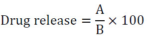
where A denotes the drug liberated from particles in milligrams, and B represents the total drug contained within the particles in milligrams.
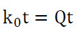
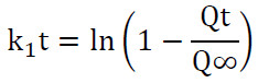
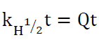
The drug-release data were analyzed using various kinetic models to clarify the release mechanism through the following equations for the
Zero-order model [45]:
First-order model [46]:
Higuchi model [47]:
And the Korsmeyer Peppas model [23,43]:
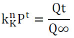
where k0 denotes the zero-order release constant, t represents time, k1 signifies the first-order release kinetics, kH refers to the Higuchi dissolution constant, P indicates the Korsmeyer–Peppas model release rate, Qt is the quantity of drug released at time t, Q∞ is the cumulative amount of drug released at infinite time, and N is the diffusional exponent that characterizes the drug-release mechanism.
RESULTS AND DISCUSSION
Particle Size, Polydispersity Index (PDI), Zeta Potential Analysis
The particle size and PDI were measured by dynamic light scattering (DLS) and the zeta potential by electrophoretic light scattering. DLS is widely used for characterization of chitosan nanoparticles and it gives the hydrodynamic particle diameter and size distribution of particles dispersed in a liquid medium [27]. Blank chitosan–TPP nanoparticles (O) showed a mean hydrodynamic diameter value of 125.5 nm with PDI of 0.190 and positive zeta potential of +25.4 mV. The results show the formation of a homogeneous dispersion with positive charged surface at nanoscale. Similar physicochemical characteristics have been reported for chitosan-TPP nanoparticles prepared by ionic gelation [27,48]. Specifically, the values of particle size, PDI and zeta potential reported by Gutiérrez-Ruíz et al. for chitosan–TPP nanoparticles prepared with 0.1% chitosan and TPP at pH 5.5 (+25.0 ± 0.79 mV; 172.8 ± 3.94 nm; 0.166 ± 0.008) confirm the physicochemical characteristics observed in the present study [48].
The particle size increased from 125.5 to 164.2 nm as a consequence of the loading of memantine (F) and the PDI increased slightly from 0.190 to 0.215. This increase could be explained by the incorporation of memantine in the chitosan-based matrix and the subsequent modifications of the intermolecular and ionic interactions that control the formation of nanoparticles. The zeta potential slightly changed from +25.4 to +21.6 mV, indicating a change of surface-charge properties after drug loading. It has been reported that entrapment of active compounds into chitosan-based nanoparticle systems resulted in changes in particle size and surface charge [27]. The particle size was altered in a concentration-dependent way by increasing B. sacra extract ratio. Formulation A, B and C showed the particle size of 169.2, 146.1 and 142.1 nm respectively with the PDI values of 0.205, 0.172 and 0.165 respectively. Hence, the extract ratio was increased to 140 μL which showed a progressive decrease in particle size and PDI showing more favorable dispersion characteristics at this concentration. The physicochemical and surface properties of polymeric nanoparticle systems can be modified by using plant-based ingredients [27].
The particle size increased 186.5 nm in formulation D and 235.2 nm in formulation E. PDI increased 0.245 and 0.312 at higher extract ratios in formulation D and E respectively. The increase in particle size and PDI at the highest extract concentration indicates increased heterogeneity and a tendency to associate or aggregate. The particle size and PDI of chitosan-TPP nanoparticles were significantly affected by the change of formulation composition and excess components [48]. The zeta potential was also affected by the concentration of the extract. The zeta potentials of the formulations O, F, A, B and C were +25.4, +21.6, +24.2, +35.8 and +41.2 mV, respectively. The observed increase in formulations B and C suggests that the addition of extract has altered the surface-charge properties of the nanoparticles. The zeta potential is an important parameter for the evaluation of the electrostatic properties and possible colloidal stability of nanoparticle dispersions [49]. Formulation C had the highest zeta potential (+41.2 mV) which correlated with higher electrostatic repulsion between the particles and may contribute to better colloidal stability. As a general rule the value of zeta potential of approx. 30 mV or more in absolute value is taken to indicate improved electrostatic stabilization. However, zeta potential is not a full indicator of colloidal stability, other factors such as ionic strength, pH and steric effects may also be involved [49]. In contrast, the zeta potential was decreased to +18.5 mV in D and +10.2 mV in E with increasing the extract ratio. The increase in particle size and PDI and decrease in zeta potential simultaneously indicates that the higher extract concentration had a negative effect on the dispersion properties and possibly increased the likelihood of particle association. Similar correlations between the surface charge and aggregation of nanoparticles have been reported in colloidal systems [50].
To summarize, formulation C showed the best physicochemical properties of the extract-loaded formulations tested, the extract of 140 μL, particle size of 142.1 nm, the lowest PDI (0.165), the highest zeta potential (+41.2 mV) and the highest transmittance (94.5 %). Therefore, formulation C was selected for further studies as the best formulation based on combined physicochemical properties and not on particle size alone. The results revealed that an appropriate concentration of B. sacra extract can positively modify the physicochemical properties of memantine-loaded chitosan-TPP nanoparticles, while excessive incorporation of extract can negatively affect the uniformity of particle size and surface charge.
Fourier Transform Infrared Spectroscopy (FTIR)
The main functional groups of the raw materials were characterized by Fourier transform infrared (FTIR) spectroscopy and the effect of the formulation on the chemical environment of the nanoparticle components was studied. FTIR spectra of prepared formulations (O, F, A, B, C, D and E) were compared to study spectral changes associated with the inclusion of memantine hydrochloride and increasing concentrations of Boswellia sacra extract. The spectrum of the blank formulation (O) showed the characteristic broad absorption region of chitosan at around 3400 cm⁻¹ due mainly to O–H and N–H stretching vibrations. The bands in the amide region and lower wavenumber region were in agreement with functional groups of chitosan and phosphate containing components. Chitosan typically presents characteristic bands at approximately 1650 and 1550 cm-1, associated with amide I and amide II vibrations, respectively, while TPP displays phosphate-related absorption bands predominantly in the 1000–1250 cm-1 range [37]. Similarly, characteristic chitosan bands and phosphate associated bands have been reported in recent studies of chitosan-TPP systems after ionic gelation [51]. The FTIR spectrum of formulation F (memantine hydrochloride) retained the main spectral features of the chitosan-TPP system. Some bands changed their position and intensity compared to formulation O indicating that the chemical environment was changed after the drug incorporation. Similar spectral shifts and change in band intensity have been reported for drug loaded chitosan nanoparticles and have been interpreted as the proof of interactions between drug and chitosan based matrix[52,53].
However, FTIR changes alone could not differentiate between drug encapsulation, adsorption or other types of association. Therefore, FTIR findings were treated as supportive evidence and interpreted in conjunction with encapsulation-efficiency results. The FTIR spectra of formulations A-E were gradually modified with increasing concentrations of B. sacra extract. The spectrum of formulation A (35 μL) was generally similar to that of formulation F with additional changes in several absorption regions. The observed changes indicate changes in the chemical environment of the chitosan-TPP-memantine system after addition of the extract, but did not remove the characteristic main spectral features. In formulation B (70 μL) changes were seen in the broad O–H/N–H region, the amide associated region and the lower wavenumber phosphate associated region. The variations could be due to the interactions between the functional groups present in chitosan, TPP, memantine hydrochloride and the phytochemical constituents of B. sacra extract. FTIR studies of chitosan–phosphate systems revealed changes in the amide and phosphate regions and were shown to be accompanied by electrostatic interactions between protonated chitosan amino groups and phosphate groups [54,55].
The changes in the spectrum were more marked in case of formulation C (140 μL) but the major features of the chitosan TPP system were preserved. The spectrum showed a broad O–H/N–H absorption band and changes in the ~2985, ~1577 and ~1074–1013 cm^(-1) regions. These alterations indicate a change in the chemical environment of the components of the formulation, after the incorporation of B. sacra extract. The persistence of the absorption features associated to phosphate suggests that the ionic network of chitosan-TPP was still present after incorporation of the extract. The ionic gelation of chitosan with TPP also showed similar retention of chitosan and phosphate related bands with a change in their position or intensity. The good FTIR characteristics of formulation C were in agreement withits physicochemical results which showed a particle size of 142.1nm, PDI of 0.165 and zeta potential of +41.2mV. These results validate the selection of the formulation C as the best formulation among the tested formulations on the basis of combined physicochemical properties. However, FTIR alone cannot be used to determine the exact localization of the extract in the nanoparticle structure or to confirm its distribution between the surface and the internal matrix.
Formulations D (280 μL) and E (560 μL) showed broad and less well resolved spectral features in a few regions at higher extract ratios. Formulation D showed changes in the O–H/N-H region and the amide- and phosphate-related regions. The observed spectral changes indicate a further change of the chemical environment with the increase of the extract concentration. The increase in particle size (186.5nm) and the decrease in zeta potential (+ 18.5mV) at the same time indicate that the higher extract concentration also affected the physicochemical properties of the dispersion. Formulation E showed the broad O-H/N-H absorption region, the variations at ~1577 and ~1507 cm-1, and the intense spectra in the low wavenumber region. There was an increase in extract concentration with particle size of 235.2nm, PDI of 0.312 and decreasing zeta potential of +10.2mV. The simultaneous changes in FTIR spectrum and physicochemical properties indicate that the highest extract concentration induced significant changes in the nanoparticle system. The results should not, however, be regarded as direct evidence for one particular mechanism of aggregation or molecular organization. The FTIR results generally indicated that the main functional-group characteristics of the chitosan–TPP system were retained after addition of memantine hydrochloride and B. sacra extract with gradual changes with increasing extract concentration. The observed changes in the amide and phosphate regions are consistent with changes in the chemical environment and interactions between the components of the formulation. The best spectral features and physicochemical properties were obtained for formulation C (140 μL) and it was selected as the best formulation for further characterization and drug-release studies.
Field Emission Scanning Electron Microscopy (FESEM)
The surface morphology and the morphological changes with increasing concentrations of Boswellia sacra extract were studied by FESEM of the dried chitosan-TPP nanoparticle formulations. The blank formulation (O) exhibited a relatively coarse and irregular morphology with aggregated structures and evident aggregation. Since the morphology is dependent on formulation and processing conditions including chitosan/TPP composition, pH and mixing conditions [31,27]. such morphological features can be observed in chitosan–TPP systems prepared by ionic gelation. The surface of memantine hydrochloride formulation F was irregular and heterogeneous with aggregated and rough structures. The drug addition had a significantly different surface appearance compared to the blank formulation. However, the FESEM images were interpreted morphologically only and were not utilized for drug encapsulation or particle size determination. Clear concentration-dependent morphological changes indicate that the increase of the extract concentration altered the organization of the chitosan–TPP matrix. Formulation A exhibited relatively smooth sheet-like regions, whereas formulation B showed more elongated fibrous or rod-like features. These differences suggest that the incorporation of increasing amounts of Boswellia sacra extract was associated with changes in the organization and aggregation of the chitosan–TPP matrix. Earlier studies incorporating Boswellia-derived constituents into chitosan/TPP nanoparticulate systems have demonstrated the compatibility of Boswellia-derived materials with this type of carrier system [56].
Of the studied formulations, Formulation C (140 μL) presented the most structured and relatively homogenous morphology with particulate structures that could be segregated within the polymeric matrix and interconnected regions could be observed. This morphology was considered the most favorable of the present series, as the individual structures were more readily discernible and less extensively aggregated than those seen at the higher concentrations of extract. In contrast, formulation D (280 μL) presented a denser, more interconnected, sponge-like morphology with less distinct individual structures. Maximum extract ratio was found in Formulation E (560 μL) with high morphological heterogeneity such as large sheet like aggregates and irregular structures. Thus, the gradual increase in the concentration of the B. sacra extract was accompanied by the characteristic morphological change from relatively separated smooth structures at low concentrations to more interconnected and aggregated structures at higher concentrations. It is well known that the modifications of the chitosan-TPP formulation conditions influence the resulting morphology and degree of aggregation [31]. The overall FESEM results showed a clear change in the morphology of chitosan-TPP nanoparticles with the addition of B. sacra extract in a concentration-dependent manner. One of the major findings of the present study is the observation of different morphological patterns for the formulations A-E. The most advantageous morphological appearance was observed in the formulation C of the investigated series. Such aggregation observed in some formulations does not necessarily indicate failure of nanoparticle formation as the FESEM analysis is representative of the dried state of the samples and the morphology could be affected by the drying process. The morphological observations obtained by FESEM should be read in conjunction with the particle size determination obtained by DLS as the two techniques provide complementary data. DLS is used to measure the hydrodynamic diameter of particles dispersed in a liquid, and the morphology of the dried specimens is revealed by FESEM. Therefore, differences between the observed morphology and DLS particle-size data may be related to hydration effects, particle aggregation, and changes during drying and sample preparation. Therefore, the morphological features observed by FESEM should not be directly considered as equivalent to the hydrodynamic particle sizes measured by DLS.
Drug Loading and Encapsulation Efficiency
Encapsulation efficiency (EE %) and drug loading (DL %) were calculated using high performance liquid chromatography (HPLC). Encapsulation efficiency (EE %) is an important parameter to evaluate the ability of a nanoparticulate carrier to retain incorporated drug during formation and isolation of nanoparticles. Drug entrapment efficiency in chitosan-TPP systems can be affected by drug properties, polymer properties, chitosan/TPP composition and the conditions employed for ionic gelation. The EE% of the memantine without Boswellia sacra extract loaded formulation (F) was 74.30% which indicates the ability of the chitosan- TPP system to encapsulate high amount of initially loaded memantine hydrochloride. Chitosan-TPP nanoparticles prepared by ionic gelation have been reported to be able to entrap high amount of drug but the EE% depends on the physicochemical properties of the drug incorporated and the formulation conditions [57]. For example, the encapsulation efficiency of the drug tacrine which was also studied for the treatment of Alzheimer’s disease was 66.1% when loaded into the chitosan–TPP nanoparticles [58].
The EE% of formulation A (35 μL) and formulation B (70 μL) were 68.80% and 70.10% respectively which were moderate reductions in comparison with formulation F with incorporation of B. sacra extract at lower ratios. These results indicate that the presence of the extract affected drug retention during nanoparticle formation. However, the specific mechanism responsible for the reduction cannot be defined by EE% alone as the changes in the composition of the formulation may affect the ionic interactions, polymer organization and drug partitioning during the formation of the nanoparticles [57]. Interestingly, formulation C among the studied formulations prepared with a ratio of chitosan to extract of 140 μL showed the highest EE% (76.80%). This value was slightly higher than that of formulation F (74.30%) and greater than that obtained for formulations A and B. The higher EE% for this intermediate concentration of extract indicates that this formulation provided better conditions for the retention of memantine in the nanoparticle matrix. This is consistent with the overall good physicochemical properties observed for formulation C before, but EE% and physicochemical properties are independent measurements and should not be taken as evidence for a specific molecular mechanism. Further increase of extract ratio decreased EE% gradually and obtained values were 64.77% for formulation D (280 μL) and 59.36% for formulation E (560 μL). The reduction at the higher extract concentrations suggests that increasing the extract amount above the 140 μL did not correlate with improved retention of memantine. Chitosan-based delivery systems have been shown to have different drug entrapment at the different formulation compositions. The amount of drug retained during nanoparticle formation can be influenced by the composition of the polymer/crosslinker and the properties of the compounds incorporated [59]. Thus, the observed trend was a decrease in EE% at low extract ratios, an increase at the intermediate 140 μL and a decrease at higher extract concentrations. Among the tested formulations, formulation C showed the highest EE% of 76.80% and formulation E showed the lowest EE% of 59.36%. Chitosan-based particulate systems containing constituents derived from Boswellia have been reported previously. For example, chitosan/TPP nanoparticles loaded with B. sacra essential oil showed an EE% of 82.7%, but the payload and formulation conditions were different from the present study [56]. In brief, EE% results showed that the concentration of B. sacra extract influenced the memantine retention in the chitosan-TPP nanoparticle system. The intermediate extract 140 μL (formulation C) showed the highest EE% and hence considered as the most favorable formulation in terms of the memantine encapsulation efficiency. The reduction at higher extract ratios indicates that higher extract concentration did not enhance drug retention. The drug loading (DL %) was determined by the percentage of the entrapped memantine hydrochloride to the total mass of the corresponding lyophilized nanoparticle formulation using Eq. [59]: DL (%) = Weight of drug entrapped Total weight of lyophilized nanoparticles × 100 DL was in the range of 6.60% to 11.62% with formulation C having the highest DL (11.62%) followed by F (10.78%), B (9.76%), A (9.55%), D (8.78%) and E (6.60%). The highest DL was observed for formulation C which was in agreement with its relatively high EE% (76.80) and lower final dry formulation mass while the lowest DL was observed for formulation E in association with its lower EE% (59.36) and higher dry formulation mass. The results overall demonstrated that the percentage of memantine remaining in the dried nanoparticle formulations was dependent on the concentration of B. sacra extract. DL% which indicates the amount of drug loaded relative to the total mass of dried nanoparticle system adds to EE%.
In Vitro Drug Release Study
The release behavior of a drug-loaded nanocarrier is dependent on its structural characteristics and the distribution of encapsulated drug in the nanoparticle matrix [60]. In Vitro Release Studies The release of memantine hydrochloride from the selected chitosan–TPP nanoparticle formulation (C) was studied in two media at 37 °C with pH values of 1.8 and 6.5. The cumulative drug release in acidic medium (pH 1.8) was 21.0% in the first 15 min, increased to 45.8% after 1 h and about 88.9% after 4 h. In the medium at pH 6.5, release was slower at the start, with 7.7% after 15 min and 19.1% after 2 h, and then rose to 92.5% after 8 h. The results showed that the release profile was dependent on the medium and time of experiment.
The difference in release profiles may be attributed to the pH-dependent properties of chitosan. Protonation of the amino groups (–NH2 → –NH3+ ) under acidic conditions may increase polymer hydration and swelling and may affect the ionic interactions between chitosan and TPP, possibly facilitating drug diffusion. The lower degree of amino-group protonation at higher pH tested may affect matrix hydration and drug transport. However, these mechanisms remain plausible explanations and cannot be confirmed from the cumulative release data alone. The release of about 88.9% of memantine within 4 h in the acidic medium indicates relatively fast drug release under the tested conditions; therefore, this result should not be interpreted as a sign of sustained or controlled release. The slower initial release at pH 6.5 indicated a different release pattern but the cumulative release was 92.5% after 8 h. Further biological studies and release-kinetics modeling are required to understand the release mechanism and to assess the potential of this formulation for drug delivery in the Alzheimer’s disease [61].
Drug Release Kinetics
The release data obtained in both media were further fitted to four established kinetic models, namely zero-order, first-order, Higuchi and Korsmeyer-Peppas, so as to further characterize the release mechanism. The corresponding rate constants and correlation coefficients (R²). The zero-order model showed the best fit (R2 = 0.9678) followed by the Higuchi model (R2 = 0.9662) in the case of the overall gastric medium. This suggested that the drug release was nearly constant and was governed mainly by diffusion. This is also consistent with the Korsmeyer–Peppas diffusional exponent (n = 0.460) as for spherical nanoparticulate carriers n is in the range of Fickian (diffusion-controlled) transport, which agrees with a matrix where acid-induced swelling dominates the release process in the absence of significant polymer erosion. A very different kinetic profile was observed in the oral medium. The Korsmeyer–Peppas model (R 2 = 0.9544) was the best fit and was superior to the zero, first and Higuchi models. The obtained diffusional exponent (n = 0.542) is in the anomalous (non-Fickian) transport region which shows that drug release in this medium is governed by a combination of diffusion through the intact polymer network and gradual relaxation or erosion of the chitosan-TPP matrix rather than by diffusion alone [62].
The combined kinetic analysis revealed that the release mechanism of formulation C was highly pH-dependent, with rapid diffusion-controlled (Fickian) release in the acidic gastric medium due to the protonation of chitosan besides the matrix swelling, while a slower anomalous transport mechanism involving diffusion and matrix relaxation dominates in the near-neutral oral medium [62].The pH-dependent release behavior of the nanoparticles is therapeutically advantageous as it suggests that the nanoparticles are able to withstand the acidic gastric environment with relatively controlled release while providing a more sustained, prolonged release profile in the near-neutral environment of the oral cavity which may facilitate the maintenance of stable memantine plasma levels and reduce dosing frequency in the management of Alzheimer’s disease .Furthermore, this pH-dependent release behavior is fully consistent with, and mechanistically explained by, the optimal physicochemical profile of formulation C previously determined by DLS, zeta potential, FTIR and FESEM characterization, further supporting its selection as the lead candidate for co-delivery of memantine and Boswellia serrata extract.
CONCLUSION
Memantine hydrochloride and Boswellia serrata extract loaded chitosan-TPP nanoparticles were successfully prepared by ionic gelation. Among all tested ratios, Formulation C (140 μL) showed the best overall performance with the smallest particle size, narrowest PDI, highest zeta potential, highest encapsulation efficiency and a favorable pH-responsive release pattern. These results confirm formulation C as a promising nanocarrier for combined memantine–Boswellia serrata delivery in the management of Alzheimer’s disease, although further biological and in vivo studies are required to confirm its therapeutic efficacy.
CONFLICT OF INTEREST
The authors declare that there is no conflict of interests regarding the publication of this manuscript.
