
Document Type : Research Paper
INTRODUCTION
Aqueous adhesive dentistry has been a fundamental tool of the modern restorative dental practice providing minimally invasive procedures while enhancing bond endurance and aesthetics with direct restorations. And, being an informational post regarding dental adhesive systems that create micromechanical and chemical retention of restorative resin composites with the dental substrates so as to increase the retention of a restoration and decrease marginal leakage. Of all adhesive systems, fifth-generation total-etch adhesives are still popular due to their clinical simplicity, bond performance and low cost. However, these adhesives continue to provide several innate limitations like incomplete polymerization, hydrolytic degradation, water sorption, Nano leakage and bacterial colonization leading to progressive deterioration of hybrid layer ultimately the failure of restoration as time elapses [1].
The stability of the polymer matrix used to formulate most dental adhesives is primarily responsible for their performance in the long-term. The methacrylate double bonds may not be fully converted during polymerization trapping unreacted monomers in the adhesive layer. These unreacted monomers impair the mechanical properties of the adhesive, promote water absorption in to it, accelerate hydrolytic degradation and even possess potential cytotoxic effects. Therefore, one of the main goals in modern dental material research was to increase the degree of conversion while keeping adequate handling characteristics [2].
Enter nanotechnology which has proved to be a rational approach to enhance the functionality of polymeric conductor-based dental materials. The use of Nano scale fillers in adhesive systems has shown great promise to improve mechanical properties, minimize polymerization shrinkage and wear, increase both thermal stability and antibacterial activity. Silica nanoparticles, titanium dioxide, zinc oxide, hydroxyapatite, bioactive glass and graphene derivatives as well as silver nanoparticles are different types of Nano materials have been studied as reinforcing agents for dental adhesives. Even with these advances, it is still a work in progress of discovering Nano materials that enhance polymerization behavior, structural integrity, surface qualities and biological performance.
According to distinct physicochemical characteristics of Cerium oxide nanoparticles (CeO₂ NPs) represented one type of nanomaterial that has received considerable attention. the fluorite structure of cerium oxide facilitated Rapid oxygen ion transport with the coexistence of Ce³⁺ and Ce4⁺ oxidation states (by doping with ceria, including oxygen vacancies). Furthermore, this unique property of reversible redox capability enables exceptional catalytic performance, chemical stability, and antioxidant effects, as well as the ability to regulate levels of reactive oxygen species. Unlike most traditional metal oxide nanoparticles, cerium oxide shows low toxicity and has high biocompatibility, making it a promising candidate for biomedical and dental applications [3].
The role of CeO₂s in resin-based adhesives may also extend to affecting the kinetics and structure of a polymer network. Nanoparticles have been shown to modify light scattering, free radical mobilization, monomer diffusion and crosslink density which in turn may affect the degree of conversion after light polymerization. These effects can either improve or impede polymer network development, depending on the concentration of introduced nanoparticles and their dispersion. In order to evaluate the interaction between cerium oxide nanoparticles and an adhesive resin matrix, it is essential to employ Fourier Transform Infrared Spectroscopy (FTIR) to evaluate polymerization both before and after curing. Nanoparticle inclusion causes microstructural surface morphology variations in cured adhesives in addition to chemical conversion. XRD shows that cerium oxide stays crystalline after being incorporated into acrylate adhesive formulations, with the exception of any new structural forms developed upon curing at elevated temperatures. SEM provides evidence of the dispersion and agglomeration states as well as interfacial compatibility between particles and matrix in the fiber reinforced polymer composites. In addition, contact angle measurements indirectly represent wettability of a surface, which plays an important role in bacterial adhesion , protein adsorption [4] and adhesive interaction/adhesion to dental tissues [5]. In addition to their catalytic behavior, cerium oxide nanoparticles also show intrinsic antimicrobial activity against both Gram-positive and Gram-negative microorganisms. Numerous mechanisms have been proposed to account for this antibacterial effect, such as oxidative stress induction [6], membrane disruption [7], interference with bacterial metabolic pathways [8] due to direct interaction of reactive oxygen species (ROS) on surfaces via chemical behavior mediated by oxygen vacancies. As secondary caries continues to be one of the leading causes of restoration failure, embedding recent generations of antibacterial nanomaterials into dental adhesives is a powerful strategy to defer bacterial colonization at the tooth-restoration interface and prolong clinical service life [9].
Although previous studies have evaluated the incorporation of different nanofillers into dental adhesive systems, there are very few comprehensive studies including physicochemical characterization, structural analysis, wettability testing, antibacterial activity and artificial intelligence-based data interpretation. Published studies generally report single properties but do not correlate the nanoparticle concentration with overall material performance in a predictable manner.
Recent developments in artificial intelligence (AI) and machine learning have ushered in a powerful new era of selective nonlinear relationship modeling capability. Thanks to the abundance of high-throughput data and machine learning, Artificial Intelligence (AI) algorithms can handle multiple experimental variables simultaneously, detect interaction between material properties that are not apparent from single-sample tests, estimate performance beyond tested conditions based on a model of readable descriptors for material properties, and optimize formulation parameters with less experiments in shorter time periods. AI has shown great potential in the fields of dental materials research, such as predicting mechanical behavior, performance of biomaterials, polymerization efficiency as well as biological response. Yet the use of this technique to smart blend formulations e.g., nanoparticle-reinforced dental adhesive formulations is hardly investigated [10].
Thus, the current study aims to evaluate the effect of different concentrations of cerium oxide nanoparticles (0; 0.25; 0.5; 1; 2 and 4 wt%) added into a commercial fifth-generation adhesive formulation. Method: The Experimental Study involved Nanoparticle size, Fourier Transform Infrared Spectroscopy before and after the light curing process, X-ray diffraction (XRD), Scanning electron microscope, Contact angle Measurement as well as antibacterial activity against Staphylococcus aureus and Escherichia coli. A machine learning-based predictive model will also be developed to determine the quantitative relationships between nanoparticle concentration and the resulting biological and physicochemical characterizations, with the aim of establishing a data-driven framework that enables future improvements to dental adhesive formulations.
Various researchers have investigated incorporating cerium oxide nanoparticles into dental biomaterials to enhance their biological and physicochemical characterizations. In this context, Byun et al. (2024) analyzed the incorporation of cerium dioxide particles into a dental adhesive to increase radiopacity; they found that CeO₂ particles could be easily incorporated without compromising the structural properties of the adhesive, while Fourier-transform infrared spectroscopy (FTIR) and X-ray diffraction (XRD) analyses demonstrated that the material‘s physical and chemical properties were adequately preserved, and also demonstrated that CeO₂ nanoparticles have the potential for use in bioactive dental restorative materials by manufactured a dental resin composite of mesoporous silica containing cerium oxide that appeared antibacterial efficacy against Streptococcus mutans without changing mechanical characterizations of the composite[11].
The results of Singh et al. (2024) showed could significantly enhance the biological performance of dental adhesive systems that indicated the cerium-containing nanomaterials exhibited effective antibacterial efficiency against oral pathogens (such as *Staphylococcus aureus* and *Lactobacillus acidophilus*), when manufactured cerium-substituted hydroxyapatite nanoparticles and incorporated them into orthodontic adhesives[6].
A way of dentistry, Zheng et al. & Chen et al. (2024) They concluded that a comprehensive review of the antibacterial mechanisms of cerium oxide nanoparticles, which linked directly between their antimicrobial performance and differents factors as particle size , the reversible redox cycle (Ce³⁺/Ce⁴⁺), oxygen vacancies , and surface chemistry[7, 10]. These distinct physicochemical properties render CeO₂ nanoparticles promising candidates for a number of biomedical and dental applications.
However, most of the published studies concerned different isolated features like antibacterial activity, radiopacity or mechanical properties[12]. There is a lack of comprehensive experiments involving the individual characterization of nanoparticles, FTIR evaluations before and after polymerization, XRD, SEM, wettability evaluation, antibacterial assessment and artificial intelligence-assisted optimization concerning CeO₂-reinforced fifth-generation dental adhesives.
This study was conducted to investigate the effect of inclusion of various concentrations (0, 0.25, 0.5, 1, 2 and 4 wt%) of cerium oxide nanoparticles into a commercially available fifth-generation total-etch dental adhesive. This was conducted on a systematic experimental basis by using particle size analysis, Fourier Transform Infrared Spectroscopy (FTIR) before and after light curing, X-ray diffraction (XRD), electron microscopic imaging methods, contact angle measurements, and evaluation of antibacterial activity against Escherichia coli. In addition, an AI-based Random Forest predictive model was established to demonstrate the quantitative relationships among CeO₂ concentration, surface wettability and antibacterial efficacy for rational prediction of optimal nanoparticle concentrations and thus the overall adhesive performances. This study advanced a comprehensive method to the synthesis of next-generation antibacterial dental adhesive systems by combining experimental properties with data-driven modeling.
This study derives its significance from combining the experimental characterization of a fifth-generation dental adhesive reinforced with cerium oxide (CeO₂) with a data-driven optimization of its composition. Rather than evaluating a single material property in isolation, this work systematically investigates the relationships associated with nanoparticle concentration—including particle dispersion, chemical and structural properties, surface wettability, and antibacterial efficacy.
Notably, an optimization strategy supported by machine learning techniques was employed to determine the cerium oxide concentration that achieves balanced, multifunctional performance, moving beyond the optimization of a single aspect alone. This structure-property-performance approach provides a rational basis for selecting nanoparticle loading levels for the development of antibacterial dental adhesive solutions.
MATERIALS AND METHODS
Materials
Methods A commercial fifth-generation light-cured total-etch dental adhesive (DENBOND TE, Vericom Co., Korea) was chosen as a resin matrix in this study. On its product data sheet, the manufacturer states that the adhesive is composed mainly of methacrylate-based monomers, hydrophilic functional monomers, photoinitiators, stabilizers and ethanol as a solvent. Materials and Methods: Cerium oxide nanoparticles (CeO₂ NPs) were used as the high purity nanofiller. We employed an appropriate particle size analyzer to determine the particle size of the nanoparticles before incorporating them into the adhesive, so that average particle diameter and size distribution could be verified.
Preparation of CeO₂-Reinforced Dental Adhesive
Experimental groups Preparation , A: Main content and The unmodified adhesive was used as the control (B), and other groups consisted of 0.25, 0.5, 1, 2 or 4 wt% CeO₂ nanoparticles respectively.
An analytical balance was used to measure the exact amounts of nanoparticles needed as aggregates, and they were added progressively into the adhesive resin. Initially each mixture was homogenized with the aid of a magnetic stirrer to allow for the first dispersion of nanoparticles into the adhesive matrix. After that, the mixes were treated by using a mechanical vibrator or vortex mixer in a lab to improve nanoparticle distribution and prevent agglomeration. The produced adhesives were preserved in opaque, light-resistant containers until characterization tests, and all mixing procedures were carried out in low ambient light to avoid premature photo-polymerization [13]. The nanoparticles of cerium oxide (CeO₂), which have been stated to be 99.9% pure, were acquired by the researcher from Ecostart Chemical (Shanghai) Co., Ltd., China. The experimental groups were determined as follows in Table 1.
Light Polymerization
The adhesive was packed into mold of definite dimensions and polymerized(LED dental curing unit) for the tested all cured specimens. With minimal distance from the specimen surface, the curing tip was arranged perpendicular to the observed phenomenon. The curing time and light intensity were kept constant for all specimens as per the recommendation of the manufacturer.
The samples were kept in the dark for 24 h after polymerization at room temperature before performing further characterization, guaranteeing a complete stabilization of post-curing [14].
Fourier Transform Infrared Spectroscopy (FTIR)
We used Fourier Transform Infrared Spectroscopy (FTIR) to analyze the chemical structure of the adhesive both before and after photopolymerization as well as to assess the effect of cerium oxide nanoparticles incorporation on the characteristic functional groups of the adhesive [15]. Fourier-transform infrared spectroscopy (FTIR) analysis was performed with a Bruker ALPHA II spectrometer. The following specimens were analyzed:
Pure CeO₂ nanoparticles
Uncured control adhesive
Cured control adhesive
Uncured 0.25 wt%
Cured 0.25 wt%
Uncured 0.5 wt%
Cured 0.5 wt%
Uncured 1 wt%
Cured 1 wt%
Uncured 2 wt%
Cured 2 wt%
Uncured 4 wt%
Cured 4 wt%
Appropriate spectral range / resolution for FTIR spectra.
Particle Size Analysis
Before being used to make adhesive, the cerium oxide nanoparticles‘ particle size was determined. Average particle diameter, particle size and polydispersity were determined to evaluate nanoparticle uniformity and investigate the possibility of forming agglomerates in the resin matrix [16]. The particle-size analysis was carried out by means of a NanoBrook 90 Plus analyzer (Brookhaven Instruments, USA).
Scanning Electron Microscopy (SEM)
Scanning Electron Microscopy (SEM) was used to examine the surfaces of all cured adhesive specimens.
Specimens were coated with a thin layer of gold using sputter coating prior to imaging in order to improve image quality and reduce the effects of charge. To evaluate the biocompatibility of materials and their microstructures, SEM micrographs at different magnifications were obtained.
Nanoparticle dispersion
Agglomeration behavior
Surface roughness
Porosity
Microstructural homogeneity
All experimental groups were compared to explore how higher CeO₂ contents affected the morphology of the cured adhesive [17].
X-ray Diffraction (XRD)
After incorporating the nanoparticles into the adhesive specimens, X-ray diffraction analysis was performed to determine what type of crystalline phases were formed. X-ray diffraction analysis (XRD) was carried out with an XRD-6000 diffractometer (Shimadzu, Japan).
Using cu-Ka radiation in an appropriate 2θ scanning range, diffraction patterns were recorded. Diffraction peaks obtained were compared with standard reference data for cerium oxide in order to confirm the crystallinity of nanoparticle and also to check whether there is any structural change after embedding ceria nanoparticles within polymer matrix.
A comparative analysis of the amorphous state of dental adhesive and the typical fluorite crystal structure of CeO2 [18].
Contact Angle Measurement
The wettability of the surface was measured through the sessile drop technique. A droplet of constant volume distilled water was carefully placed on the as-prepared adhesive surface, and the static contact angle was measured with a digital contact angle analyzer. For each specimen, three measurements were taken and averaged. The contact angle variations were interpreted in terms of the influence of CeO₂ nanoparticles applied on [19]:
Surface energy
Hydrophobicity
Potential bacterial adhesion
Clinical wetting behavior
Antibacterial Activity
The antibacterial activity of the cured adhesive specimens was tested against two types of bacteria obtain from Environmental Research Centre - University of Technology :
Staphylococcus aureus (Gram-positive)
Escherichia coli (Gram-negative)
Microbiological Method (Antibacterial Activity when it occurs agar diffusion) Each group was incubated under a standard condition (37˚C for 24h) in nutrient agar for each bacterial culture, then diluted to obtain concentration ( 1011 CFU/ ml) , then the antibacterial activity of every group was measured according to [20], where 100µ of each inoculum for each type of bacteria spread on mular-Hinton agar then CeO₂-containing and control adhesive formulations put on this agar. The inhibitory efficiencies of CeO₂-containing and control adhesive formulations were evaluated in order to assess the impact of nanoparticle dosage on antibacterial efficacy by measured the diameter of inhibition zone (mm) [21].
Statistical Analysis
The mean ± standard deviation (SD) of three separate, triplicate experiments is used to express the results. To find significant differences between the experimental groups, statistical analysis was carried out using one-way ANOVA and Tukey‘s honestly significant difference (HSD) post hoc test. The definition of statistical significance was p < 0.05. Python (SciPy and Statsmodels packages) was used for all statistical calculations [22,23].
AI-Based Modeling
To computationally determine predictive correlations between the concentration of cerium oxide nanoparticles and the regime-appropriate physicochemical and biological performance of the adhesive, an AI-based model was created. Contact angle and antibacterial activity against E. coli were the experimentally determined performance parameters for the data-driven analysis, while the CeO₂ nanoparticle concentration was the input variable in the experimental dataset.
Furthermore, the contribution of each experimental variable to the material‘s predicted performance will be determined, a well-known method in the field of Explainable AI (XAI). This technique provides a quantitative approach to identifying the optimal CeO₂ concentration, enabling the simultaneous enhancement of surface properties, antibacterial activity, and polymerization efficiency.
RESULTS AND DISCUSSION
Particle Size Distribution of Cerium Oxide Nanoparticles
Between around 45 and 70 nm, majority of particles were concentrated, with very few observed in the lower and higher size ranges. The particle size distribution exhibited a unimodal pattern; there was no indication of excessively coarse particles, and the size distribution curve closely resembled a normal distribution, indicating a high degree of uniformity among the particles and the average nanoparticle dimension was 58.22 nm, confirming that the reinforcing particles fall within the nanoscale range. The size distribution of the cerium oxide (CeO₂) nanoparticles used in this study will illustrates in Fig. 1, The dimensions of the CeO₂ nanoparticles provide a large surface area, which is expected to facilitate intimate interaction with the adhesive resin matrix following incorporation,. Furthermore, the observed particle sizes suggest suitability for dispersion within dental adhesives, enabling subsequent mechanical and physicochemical testing [24].
Fourier Transform Infrared Spectroscopy (FTIR)
Figs. 2–4 Present FTIR spectra of pure CeO₂ nanoparticles, control adhesive and CeO₂-modified dental adhesives before or after light curing. The similar spectral characteristics of all experimental formulations suggested that the addition of cerium oxide nanoparticles did not change the basic chemical structure of fifth-generation dental adhesive.
The unmodified adhesive had the spectrum of a methacrylate-based resin system with attendant characteristic absorption bands. Observing a strong absorption peak for ester carbonyl (C=O) groups stretching vibration around 1710–1714 cm⁻¹. A second characteristic absorption band around 1635–1638 cm⁻¹ was attributed to the stretching vibration of the C=C in methacrylate monomers, indicating a reactive functional group that participates in free-radical polymerization. Other group absorption bands in the 1290–1040 cm⁻¹ region were assigned to C–O–C stretching vibrations of ester linkages, and some peaks below 900 cm⁻¹ were attributed to skeletal vibrations of the polymer network [25].
Absorption bands were not further observed in the spectral range studied following addition of CeO₂ nanoparticles at concentrations 0.25, 0.5, 1, 2 and 4 wt% (corresponding to molar volume fraction Xmol %/g=14%) [14]. Similarly, as for lateral shift into the respective regions of principal functional groups identified, no new detectable chemical bondings between added nanoparticles and resin components can be evidenced. All modified adhesive formulations retained the carbonyl absorption band at ~1712 cm⁻¹ after light curing (Fig. 3a), indicating preservation of the polymer backbone during photo-polymerization. Oppositely, the absorption intensity around 1636 cm⁻¹ due to the aliphatic methacrylate double bond was diminished in the cured specimens compared with their corresponding uncured formulations pointing towards progressive consumption of reactive double bonds during polymer network formation.
The fingerprint region (around 1300–800 cm⁻¹) remained relatively unchanged for all nanoparticle concentrations, albeit with slight peak intensity differences between experimental groups. The small differences are related to alterations of polymer crosslinking density and due to dispersed CeO2 nanoparticles, not the creation of chemical species [2].
For all concentrations of the nanoparticles (0.25–4 wt%), it was observed that increasing the CeO₂ content did not noticeably change the position of characteristic functional groups compared to pure material. Rather, differences observed among formulations were due to changes in the peak intensity before and after photo-curing, indicating that the primary effect of CeO₂ nanoparticles is related to their influence on polymerization behavior/network development rather than any inherent change in the chemistry of the adhesive.
In summary, FTIR analysis clearly showed that CeO2 NPs were integrated appropriately into the adhesive matrix carrying the fundamental chemical structure of the same original resin system. The decrease in the aliphatic C=C absorption band as observed in light cured samples indicates polymerization of all experimental formulations [26].
X-ray Diffraction (XRD)
Crystal Structure of Cerium Oxide Nanoparticles
The pristine cerium oxide (CeO₂) nanoparticles X-ray diffraction (XRD) pattern is shown in Fig. 5. The diffraction patterns showed sharp crystalline reflections representing cerium oxide with a fluorite structure and suggesting the high crystallinity of these nanoparticles synthesized.
The strongest diffraction peak was located at around 2θ = 28.6°, which could be assigned to the (111) crystal plane. As shown in Fig. 2b, diffraction peaks are found to be in good agreement with the standard reference pattern of cubic fluorite CeO₂ (ICCD card No. 03-065-2975) apart from confirming that phase pure cerium oxide nanoparticles have been formed successfully [27].
No diffraction peaks resulting from secondary cerium oxide phases or impurity phases were observed in the scanned diffraction range indicating that the nanoparticles contained a very high phase purity
This is further validated by the narrow and intense diffraction peaks indicating an inherently crystalline nature of CeO₂ nanoparticles as it was anticipated that no structural changes were observed in the individual nanoparticles before being blended into a dental adhesive matrix [28].
Structural Analysis of CeO₂-Reinforced Dental Adhesives
The X-ray diffraction patterns for the unmodified (Control) and CeO₂-reinforced adhesive formulations of 0.25, 0.5, 1, 2 and 4 wt% nanoparticles are shown in Fig. 6
The unmodified adhesive diffractograms yielded a wide diffuse halo centered at about 2θ = 20°, indicating that the polymeric resin matrix was almost entirely amorphous. Due to the crosslinked methacrylate-based nature of the dental adhesive, a no sharp crystalline diffraction peaks were obtained.
The broad amorphous halo stayed to dominate the structure after addition of CeO₂ nanoparticles, and the experimental materials appeared a comparable amorphous diffraction pattern regardless of their concentration which indicating that cerium oxide nanoparticles had minimal impact on the polymer matrix‘s overall structural organization [29].
Fig. 5 appeared prominent diffraction peaks associated with the cubic fluorite (Fm3m) crystal structure resulting from The crystallized CeO₂, while the modified adhesive samples did not clearly reveal these distinctive reflections when used the XRD patterns, as well as the lack of discernible CeO₂ diffraction peaks which may account result from low nanoparticle loading, their uniform distribution within the amorphous polymer matrix, and the resin phase‘s dominant contribution to scattering, which obscures weak crystalline reflections originating in nanoparticles.
No impurity peaks or additional crystalline phases were detected across the examined diffraction range.
Overall, these results indicate that the amorphous nature of the dental adhesive was largely preserved due to the addition of cerium oxide (CeO₂) nanoparticles. However, the pristine CeO₂ showed its distinctive cubic fluorite crystalline structure, and the addition of CeO₂ nanoparticles did not change the adhesive matrix‘s predominantly amorphous character [30].
Scanning Electron Microscopy (SEM)
Scanning electron microscopy (SEM) was used to monitor the surface morphologies of cured control adhesive and CeO₂-reinforced adhesive formulations for each group; typical SEM micrographs are displayed in Fig. 7. With 394 isolated surface imperfections, the control adhesive (Pad) exhibited the smoothest and most uniform surface (Fig. 7a). The resulting polymer matrix during photo-polymerization is demonstrated by the absence of particle features. Different nanoparticle domains were introduced into the resin matrix by adding 0.25 weight percent CeO₂ nanoparticles (Fig. 7b). Because cerium oxide nanoparticles tend to clump because of their high surface energy, the particles were found to be pretty well scattered with a few minor agglomerates. This was for glue containing 0.5 weight percent CeO₂ (Fig. 7c), which was comparatively homogenous with little particle agglomeration. The uniformly distributed particles show good mixing and compatibility with the adhesive glue since they stay entrenched in the polymer matrix without causing noticeable surface flaws [31]. Larger irregular aggregates were also seen dispersed across the surface when we raised the nanoparticle concentration to 1 weight percent (Fig. 7d). clusters in this area indicate partial agglomeration, resulting from the inter particle distance decreases with increasing filler loading. A greater number of agglomerated particles with larger aggregate diameters was observed when the CeO₂ concentration was increased from 1 wt% to 2 wt% (Fig. 7E). The dispersion of the nanoparticles was less uniform compared to the lower-concentration case, although they remained present within the cured adhesive. At the greatest concentration (4 wt%, Fig. 7f) Particle agglomeration was present which resulting from the volume of resin available was inadequate to properly separate the closest neighboring nanoparticles and resulted in secondary particle clustering and formation of irregular, micron-sized clusters dispersed throughout the resin matrix [32].
The dispersion of CeO₂ nanoparticles inside the adhesive matrix and surface shape are qualitatively shown by the SEM pictures. The micrographs displayed a comparatively homogenous morphology with little discernible aggregation at lower CeO₂ concentrations (0.25 and 0.5 wt%). More noticeable irregular aggregates were seen as the CeO₂ concentration increased, especially at 1, 2, and 4 weight percent. These findings point to a concentration-dependent tendency toward particle agglomeration; however, the images were not used to quantitatively characterize the degree of aggregation or determine an exact particle-size distribution because the SEM analysis in this study was primarily qualitative. Therefore, rather than quantitative particle-size measurements, the SEM results are primarily interpreted in terms of overall morphological changes and apparent dispersion patterns, These findings characterize the cured adhesive‘s surface morphology; they do not, by themselves, show modifications in wettability, polymerization behavior, or antibacterial efficacy. The observed morphological variations are therefore considered as possible correlations rather than proof of a causal process, and the data pertaining to contact angle and antibacterial effectiveness are analyzed separately [33].
Wettability Analysis
The static water contact angle (SWCA) was performed to evaluate the surface wettability of cured dental adhesive formulations, and the results are listed in Fig. 8 and Table 3.
Bond: The unmodified adhesive showed a contact angles of 66.8 ± 1.6° which demonstrated the intrinsic wettability nature of commercial fifth-generation dental adhesive After CeO₂ nanoparticle being introduced, a gradual decrease in contact angle was obtained with an increase of the particle dosage from 0.25 wt% to 1 wt%.
The contact angle decreased from 62.9 ± 1.4° for the 0.25 wt% formulation to 58.7 ± 1.3° at the concentration level of strictly disaggregating CeO₂ (0.5 wt%), dropping further to a minimum value of ≃54.8 ± 1.5° at the highest concentration level used (1 wt% CeO₂) This gradual decrease signifies that the surface wettability is significantly improved by the incorporation of nanomaterials [34].
An increase in nanoparticle concentration to 2 wt% gave a relatively slight [in comparison with values of the control adhesive] increase in contact angle - up to 57.6 ± 1.8 °, but for the highest (4 wt%) the contact angle was 63.5 ± 2.0 ° close to but lower than that obtained on the surface of control adhesive.
In summary, the wettability data demonstrate a concentration dependence; low to moderate concentrations of CeO₂ significantly enhanced the hydrophilicity of the adhesive surface, whereas higher nanoparticle concentrations partially negated this effect.
Furthermore, this pattern aligns with scanning electron microscopy results. At low concentrations, the uniformly dispersed cerium oxide nanoparticles increased the effective surface energy for adhesive solidification at the interface, thereby enhancing wettability. Conversely, at higher concentrations (2–4 wt%), larger nanoparticle aggregates produced a heterogeneous surface morphology, leading to a partial recovery of the contact angle.
Among the studied groups, the (1 wt%) CeO₂ formulation showed the lowest contact angle, indicating the highest surface wettability. Nevertheless, enhanced bond strength or interfacial adaptation to oral tissues cannot be established by contact-angle measures alone. These results necessitate specific adhesive bond-strength testing and additional assessment in clinically relevant settings [35].
One-way ANOVA of the data (contact angle) showed a highly significant effect of CeO2 concentration on surface wettability (F(5,12)=67.82, p<0.001). Tukey‘s post hoc test showed that the 1 wt% formulation had the lowest contact angle and was significantly different from all other groups (p < 0.05). The formulations containing 0.25 wt% and 4 wt%, as well as those with 0.5 wt% and 2 wt% respectively did not differ significantly from one another. These results corroborate that additions of ~1 wt% CeO2 lead to the most effective enhancement of adhesive wettability.
Antibacterial Activity of the cured CeO₂-Reinforced Dental Adhesive
Then, the antibacterial activity of the cured polymeric formulations was evaluated against one Gram negative and another Gram positive bacteria (Escherichia coli and Staphylococcus aureus, respectively) by over-time agar well diffusion assay. Table 4 and Fig. 9 present the inhibition zone diameters
No antibacterial activity was obtained against E. coli for the control adhesive (Bond) having a diameter of inhibition zone equal to 0 mm. After the addition of CeO₂ nanoparticles, antibacterial results were significantly enhanced. The 0.25 wt% formulation created a 10.0 mm inhibition zone, while the zonal diameters were slightly larger at 18.0 mm and 18.3 mm for two subsequent formulations tested at concentrations of both 0.5 and 1 wt% (Authors own data). The inhibition zone area of the 2 wt% formulation showed modestly low (17.0 mm) when compared to the others whereas, The greatest antibacterial activity obtained was for the 4 wt% formulation that produced an inhibition zone of 19.0 mm.
A generally increasing trend in antibacterial activity was seen with increasing CeO₂ concentration, however the improvement did not follow a straight linear increase. The diameter of the inhibition zone decreased somewhat at 2 weight percent compared to 1 weight percent before expanding once again at 4 weight percent; this indicated that the concentration of nanoparticles had an impact on antibacterial activity [36].
The antibacterial effect of the CeO₂-containing adhesive was dependent on the type of microorganism, as indicated by the absence of a visible inhibition zone against S. aureus bacteria under the specified experimental conditions. This result emphasizes that antibacterial efficacy cannot be applied to all bacterial species and may rely on the properties of the bacterial cell envelope, interactions between nanoparticles and bacteria, and the capacity of active species to diffuse from the adhesive matrix [20].
In summary, the obtained results indicate that introducing up to 2 wt% of CeO₂ nanoparticles into an cured dental adhesive has a significant effect on its antibacterial properties against E. coli, with no inhibitory effect found against S. aureus within the concentration range studied here [37].
Antibacterial activity against Escherichia coli was most significantly related to CeO₂ concentration (One-way ANOVA, F(5,12) = 5305.42, p 0.05), suggesting that increasing nanoparticle content above a threshold value of only limited further increases in antibacterial efficacy.
Adhesive performance does not simply improve monotonically with increasing CeO₂ concentration. Rather, resulting an ideal concentration that equilibrium of antibacterial activity, surface wettability, and nanoparticle dispersion.
AI-Based Modeling and Optimization
AI-assisted modeling is considered an attractive strategy to expedite the development of advanced dental biomaterials in terms of identifying favourable material compositions with minimal experimental trials. MPI enables machine learning algorithms which can build missing parts of the solution space by capturing complex nonlinear relations between formulation parameters and multiple material properties in a single step, rather than traditional statistical analysis. The objective of the present study was to establish a predictive framework using artificial intelligence (AI) tools, in order to assess the impact of varying concentrations of cerium oxide (CeO₂) nanoparticles on the performance of an experimental dental adhesive.
A Random Forest Regression (RFR) algorithm was chosen due to its superior feature of providing coverage for non-linear datasets without assuming any existing mathematical relations between features. The model was trained on experimentally measured concentrations of CeO₂ nanoparticles in addition to the corresponding wettability (contact angle) and antibacterial activity against Escherichia coli. The functional properties of the adhesive which were experimentally measured were integrated in a single normalized performance item representing the Composite Performance Index (CPI), allowing simultaneous optimization of two major functions of an adhesive.
In contrast to traditional optimization methods which necessitate numerous exploratory formulations, the AI model provides predictions on material performance across a continuous concentration spectrum and highlights the composition that yields optimal properties. This method not only offers fresh insights on formulation creation, but it also significantly reduces the load of experimental work [38].
Creating the Random Forest Model
The association between CeO₂ concentration and the composite performance indicator was investigated using the Random Forest regression model as an exploratory tool. Using model-based interpolation, predictions were produced at 300 concentration sites within the studied range. These points do not constitute further independent experimental findings; rather, they are computational predictions. The results are considered exploratory due to the small experimental dataset, and further formulations and independent experimental measurements are needed for validation.
We used Scikit-learn ML python library to implement Random Forest Regression model. The independent input variable was taken as the concentration (wt%) of CeO₂, while the experimentally determined wettability and antibacterial activity were combined into a single performance descriptor through normalization.
Normalized contact angle values were calculated such that lower contact angles would produce a higher normalized score, which reflects a more desirable level of wettability (contact angle). In the same way, measured diameters of the inhibition zones from E. coli antibacterial assay were normalized so that more effective inhibition zones corresponded to higher scores on antibacterial activity (Fig. 2b).
Composite Performance Index composite was then calculated in accordance with [39]:
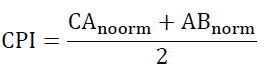
Where, CAnorm represents the normalized wettability score, ABnorm represents the normalized antibacterial score.
The Random Forest model was trained with 500 trees with a fixed random seed, for reproducibility. Once trained, the model provided predictions on the composite performance in-between 0 and 4 wt% CeO₂ for a total of 300 concentration points (interpolated) yielding a continuous prediction across that range.
The CPI (Composite Performance Index) was computed by summing up the normalized values of both the Contact Angle and Antibacterial Activity, and giving each equal weight:
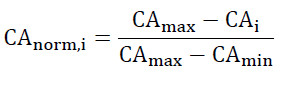
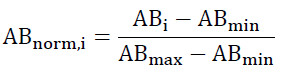

The contact angle (CA_i) of formulation (i) and the inhibition zone diameter (AB_i) of formulation (i) against E. coli are both represented by this equation. The minimum and maximum limits of each parameter have been established using the experimental data set. Formulations with low contact angles received higher normalized scores. Formulations with large inhibition zones received higher antibacterial activity scores. A weighting factor of 0.5 was applied to each objective as both wettability and antibacterial activity are equally important objectives when optimizing a formulation. Thus, the CPI ranges from 0 to 1, with the higher value representing a better balance between the two selected objectives.
AI Prediction Results and Discussion
The Random Forest model was able to capture the non-linear relationship of CeO₂ concentrations to overall functional performance of experimental dental adhesive. After the onset of CeO₂ nanoparticle, as seen in Fig. 10, an obvious rise was observed for the predicted Composite Performance Index and this indicates that minor additions of nanoparticles may be associated with a substantial enhancement of material performance.
The peak in the prediction matched closely with an approximately 1 wt% CeO₂, which occurs at nearly the same composition of one of the lowest contact angles (along with one of highest antibacterial activities against E. coli) in terms of experimental formulation measured. The Random Forest model identified 1 wt% CeO₂ as the formulation providing the highest composite performance within the investigated concentration range, and this concordance between the AI predictions and laboratory observations.
This indicated that the addition of 1 wt% may be associated with an optimum performance, above which the predicted performance started to decline slowly, even if antibacterial activity continue to rise. At higher concentrations of nanoparticles, both viscoelastic properties and wettability deteriorated concurrently which might be attributed to excessive loading of CeO₂ boosting the agglomeration along with a diminished dispersion in the adhesive matrix. Consequently, overall performance is achieved by balancing antibacterial efficacy and surface wettability, rather than relying on a single property,
the expected optimal standard points to a formulation that achieves an ideal balance between the two factors, interfacial biological activity and behavior because equally important for ensuring strong adhesive performance in restorative dentistry.
Feature Importance Analysis
While only a small selection of experimental variables were included in the current work, the Random Forest algorithm offers an insight into how influential each property that was measured is to generating performance composite.
Derivation of the composite performance index using the multivariate regression model showed that wettability and antibacterial activity are major contributors in the prediction. The lower effect of the wetting angle on the optimization was due to its progressive improvement from control sample to 1 wt% formulation, followed by an increase in larger concentrations of CeO₂. For example, the antibacterial activity against E. coli generally increased almost monotonically as loading of nanoparticles was raised, leaving smaller differences for the highest concentrations.
This makes sense as a method, since these AI models seemed to associate surface wettability with the driving factor for determining optimal formulation while antibacterial performance was identified more as a property that plays a supportive role. Consequently, rather than optimizing each of them in isolation, the best overall performance is achieved by balancing these two attributes simultaneously [40].
Experimental Characterization vs AI Prediction
AI-driven materials design offers a key advantage: the integration of characterization data from multiple sources into a unified predictive framework. The concentration that performed the best among the formulations examined under the particular standards assessed in this investigation predicted by the Random Forest model—1 wt% CeO₂—aligns closely with the experimental observations made in this study.
Fourier-transform infrared (FTIR) spectra revealed no new chemical functional groups or changes in the polymerization mechanism of the adhesive system upon the incorporation of cerium oxide nanoparticles. Instead, only slight fluctuations in peak intensity were observed, indicating that the nanoparticles were successfully incorporated into the polymer network without causing significant disruption.
Similarly, following the incorporation of the nanoparticles , this cerium oxide (CeO₂) retained its characteristic fluorite crystal structure, nor the binding material (matrix) underwent no significant change, preserving its initial amorphous nature, This highlights the chemical stability of the nanocomposite, as no other crystalline phases were detected when examined withX-ray diffraction (XRD) analysis.
The AI prediction was also corroborated by SEM observed surface morphology. In contrast to higher nanoparticle concentrations, which demonstrated increasingly larger levels of clustering, the formulation for 1 weight percent displayed a rather uniform dispersion of the CeO2 nanoparticles with few agglomerates. This adhesive aggregation is the adhesive polymer matrix.
These observations were further corroborated by the wettability measurements. The surface contact angle decreased gradually from 66.8° for the unmodified adhesive to 54.8° at 1 wt% CeO₂, proving the great increase in hydrophilicity of the adhensive surface. Above this value, the contact angle increased steadily when the nanoparticle loading was increased, indicating a gradual loss of surface homogeneity possibly due to excess nanoparticles.
From this, it could be seen that with the increase of CeO₂ concentration, there was a higher inhibition ability to E. coli, yet the improvement seems moderate after 1 wt%. As a result of this, the AI algorithm appropriately recognized that an increase in nanoparticle concentration is not always advantageous for improvement of overall material performance due to compensatory changes—improvements in antibacterial activity will reach a plateau as wettability is decreased.
Hence, the remarkable agreement between actual and predicted values for FTIR, XRD, SEM, wettability and antibacterial behaviours indicate that machine-learning approaches can successfully predict optimal nanocomposite formulations using relatively small experimental datasets.
Practical Implications
Some practical advantages of the proposed AI-assisted optimization strategy for designing novel multifunctional dental adhesives are discussed.
• It significantly cuts down the number of experimental formulations necessary while optimizing material.
• It allows several material properties to be considered at once, instead of optimizing one property at a time.
• Rapid prediction of optimum nanoparticle concentrations prior to large scale laboratory experiments
• Costly materials, experimental work and development time are reduced.
• The designed framework is easily expandable to other experimental variables (e.g., the shear bond strength, degree of conversion, microhardness at distinct timepoints and volumetric water sorption—water uptake or cytotoxicity).
• The methodology can be easily integrated by modern Materials Informatics approaches for the next-generation dental biomaterials.
Future AI Applications
The current study used Random Forest Regression which is robust with small datasets, however future studies could benefit from more advanced machine-learning approaches to better predictive performance.
Potential future approaches include:
Artificial Neural Networks (ANN)
Extreme Gradient Boosting (XGBoost)
Gaussian Process Regression (GPR)
Support Vector Regression (SVR)
Bayesian Optimization
Multi-objective evolutionary optimization algorithms
In addition, the deep-learning image analysis could be wider to integrate with SEM micrographs for automatic quantification of nanoparticle agglomeration, particle distribution, porosity and surface defects. These image-derived descriptors could then be paired with conventional physicochemical measurements to define fully automated structure–property–performance relationships for dental nanocomposites.
Summary of AI Modeling
The Random Forest model successfully discovered the non-linear association between CeO₂ nanoparticle concentration and aggregate functional performance of the experimental dental adhesive. This study finded, that experimental may be associated with both wettability and antibacterial activity tests, showed aligned remarkably well with AI predictions indicated that a 1 wt% CeO₂ content represents the optimal composition.
The close match between experimental characterization and machine-learning-prediction illustrates that AI-assisted optimization is a powerful complementary tool for the rational design of dental nanocomposite adhesives. Instead of substituting laboratory experimentation, the proposed approach efficiently navigates formulation development by concentrating on high promise compositions and minimizes testing non-viable formulations.
Study Limitations
When evaluating the results of this study, a number of limitations should be taken into account.
First, as the study was carried out in vitro, it is not possible to immediately apply the findings to clinical performance.
Second, the model should be considered exploratory and its predictions need to be validated using additional formulations and independent datasets because the machine-learning analysis was based on six experimentally tested CeO₂ values.
Third, Streptococcus mutans, a significant oral cariogenic microbe, was not examined; instead, the antibacterial evaluation was restricted to Escherichia coli and Staphylococcus aureus. Furthermore, water-sorption measurements, quantitative degree-of-conversion analysis, bond-strength testing, long-term aging and durability evaluation, and cytotoxicity testing were not carried out. As a result, the current findings do not prove biological safety, clinical bonding performance, or long-term adhesive durability. therefore Before the clinical application of CeO₂-reinforced dental adhesives can be determined, more research including these evaluations is required.
CONCLUSION
However, within the design constraints of this study, it can be concluded that CeO₂ nanoparticles were effectively incorporated into a commercial fifth-generation dental adhesive while maintaining its core chemical structure (FTIR verified). The incorporation of CeO₂ (0, 0.25, 0.5, 1, 2, and 4 wt%) did not change the crystal structure of the CeO₂ but changes in adhesive results showed that while crystalline size slightly decreases with increasing amounts of CeO₂, the amorphe content remained dominant as well as similar sizes. Low loading ratios (0.25–1 wt%) of CeO₂ have not significantly affected the homogeneity of structural properties in our sample materials, as suggested by SEM observations compared to samples containing higher amounts that led to excessive agglomeration and reduced dispersion homogeneity between particles. With the addition of CeO₂ nanoparticles, a significant increase in adhesive surface wettability was observed and the contact angle value obtained with water (54.8°) at 1 wt% CeO₂ showed that it reached the best surface characteristics among all tested formulations. Nanoparticle incorporation enhanced antibacterial activity against Escherichia coli, achieving the greatest inhibition zone (19 mm) with the 4 wt% formulation, while no detectable antibacterial activity against Staphylococcus aureus was observed under the current experimental conditions. In addition, the nonlinear relationship of the overall material performance versus nanoparticle concentration have been well described by the AI-assisted Random Forest modeling. It has been shown that an improvement on the wettability and antibacterial activity is obtained, and According to the chosen Composite Performance Index, among the six formulations that were experimentally studied, 1 weight percent CeO₂ offered the best balance between contact angle and antibacterial activity. Before more general inferences can be made, more experimental validation is necessary because this finding is unique to the tested formulations and evaluation criteria. The good agreement between experimental results and AI predictions illustrates that machine-learning-assisted optimization is a viable pathway to accelerating dental nanocomposite adhesive design while minimizing both the quantity of experiments performed and materials consumed. In general, the proposed methods provide a promising strategy for future optimization of dental adhesive compositions using nanotechnology and experimental characterization together with artificial intelligence.
CONFLICT OF INTEREST
The authors declare that there is no conflict of interests regarding the publication of this manuscript.
